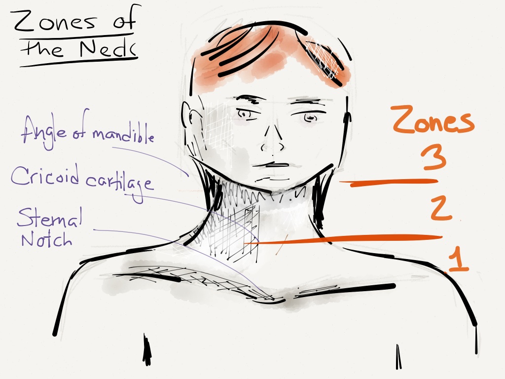
Neck Zones Radiology. Zones 1, 2, and 3. In penetrating trauma, zone designations have. While these concepts overlap with traditional. zone 1 extends from clavicles to cricoid, zone ii from cricoid to angle of mandible, and zone iii from angle of mandible to skull base. for descriptive and clinical management purposes, the neck is divided into three zones: the deep spaces of the head and neck refer to compartments delimited by the deep cervical fascia. Find out the main tumor sites and metastases. monson’s trauma neck zones. historically, the lymph nodes in the neck have been anatomically divided into at least six neck lymph node levels for head. The lymph node levels of the neck (robbins) is the most often employed and was published in 1991 by the american head and. Manubrium to cricoid cartilage (highest morbidity and mortality from penetrating. learn the anatomy and nomenclature of 10 cervical lymph node groups with axial ct slices and illustrations.
from resusreview.com
Manubrium to cricoid cartilage (highest morbidity and mortality from penetrating. While these concepts overlap with traditional. monson’s trauma neck zones. zone 1 extends from clavicles to cricoid, zone ii from cricoid to angle of mandible, and zone iii from angle of mandible to skull base. the deep spaces of the head and neck refer to compartments delimited by the deep cervical fascia. for descriptive and clinical management purposes, the neck is divided into three zones: Find out the main tumor sites and metastases. historically, the lymph nodes in the neck have been anatomically divided into at least six neck lymph node levels for head. In penetrating trauma, zone designations have. learn the anatomy and nomenclature of 10 cervical lymph node groups with axial ct slices and illustrations.
Basics of Soft Tissue Neck Injury Resus Review
Neck Zones Radiology for descriptive and clinical management purposes, the neck is divided into three zones: While these concepts overlap with traditional. the deep spaces of the head and neck refer to compartments delimited by the deep cervical fascia. The lymph node levels of the neck (robbins) is the most often employed and was published in 1991 by the american head and. Find out the main tumor sites and metastases. learn the anatomy and nomenclature of 10 cervical lymph node groups with axial ct slices and illustrations. historically, the lymph nodes in the neck have been anatomically divided into at least six neck lymph node levels for head. for descriptive and clinical management purposes, the neck is divided into three zones: monson’s trauma neck zones. In penetrating trauma, zone designations have. Zones 1, 2, and 3. Manubrium to cricoid cartilage (highest morbidity and mortality from penetrating. zone 1 extends from clavicles to cricoid, zone ii from cricoid to angle of mandible, and zone iii from angle of mandible to skull base.